Keratoconus treatment
Keratoconus is a gradual thinning and «bulging» of the cornea, which leads to image distortion, glare, and reduced visual acuity.
Keratoconus treatment with the BritishX method – first results in 12 minutes
Contact us
If you have any questions, call our friendly call center or make an appointment for diagnostics with further consultation.
Mon - Sun 9:00 – 19:00
we work without days off
Keratoconus treatment with the BritishX method – first results in 12 minutes
Keratoconus is a progressive corneal disease in which the cornea becomes thinner and deformed, taking on a conical shape. The condition is not inflammatory but affects vision. The main risk is a significant decrease in visual acuity, which can deprive you of the ability to drive, work with text, or perform everyday visual tasks.
Among the symptoms of keratoconus are distorted vision, increased light sensitivity, burning or itching in the eyes, and double vision. These manifestations interfere with normal sight, reduce concentration, and significantly impact quality of life.
What happens if treatment is ignored?
What happens if treatment is ignored?
The disease usually develops gradually and leads to severe corneal changes. Possible complications include:
- Development of severe astigmatism;
- Persistent double vision (diplopia);
- Corneal edema (hydrops);
- Corneal ruptures;
Without timely intervention, the structural changes in the cornea become irreversible. Advanced keratoconus may result in complete vision loss, especially if effective measures are not taken early. Only early diagnosis and properly selected therapy can slow down or completely stop disease progression.
What causes keratoconus?
What causes keratoconus?
The development of keratoconus is associated with dysfunction in the brain’s regulatory functions. All body processes, including vision, are controlled by the central nervous system. When neural connections and the interaction of visual control centers are disrupted by internal or external factors, system imbalances begin to occur.
Like many chronic diseases, keratoconus develops due to a combination of factors: genetic predisposition, visual strain, eye trauma, hormonal imbalance, immune disorders, and others. Together, these influences can destabilize the visual system and lead to characteristic corneal changes.

Take care of your vision health today!
Book an eye examination to learn more about the condition of your visual system!
Mon - Sun 9:00 – 19:00
we work without days off
How to effectively stop the progression of keratoconus?
How to effectively stop the progression of keratoconus?
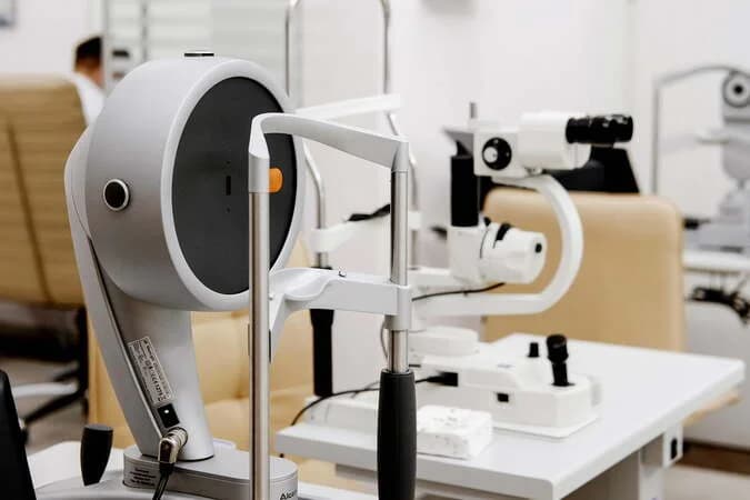
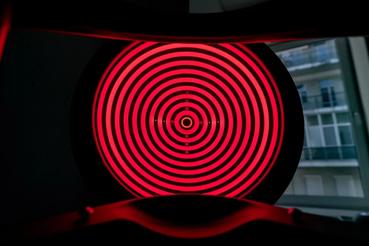
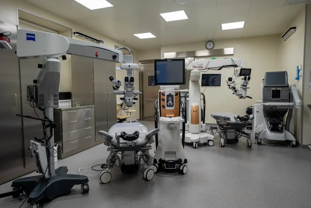
Modern ophthalmology offers an advanced, proven, and safe method for treating keratoconus — the cross-linking procedure. This technology strengthens the corneal tissue by creating additional bonds between collagen molecules. The approach is especially relevant for patients under the age of 45, when the disease tends to progress most actively.
The method was developed in Switzerland and is based on targeted ultraviolet exposure to the cornea, which is pre-treated with riboflavin. The controlled light penetration depth activates biochemical processes that make the tissue denser and more resistant to deformation. Disease progression is halted, and over time, stabilization or even partial recovery of visual function may occur.
Modern cross-linking: guaranteed results
Modern cross-linking: guaranteed results
We also offer an advanced cross-linking technique for stabilizing the cornea at the early stages of keratoconus. The cost of the procedure depends on the chosen protocol and diagnostic parameters. You can clarify the cross-linking price during your consultation.
We use only certified equipment and Swiss-made pharmaceuticals. The cross-linking procedure helps preserve corneal health and prevent further disease progression, especially when keratoconus is detected early. If you’re interested in the cost of cross-linking, our consultants will provide full information tailored to your specific case.
doctors
3
specialized doctors
Experts in keratoconus treatment
Our doctors will carefully study the condition of your visual system and determine whether correction is possible in your case.

Experience since 2012
Zhykharev Anatolii
Ophthalmic surgeon, refractive surgeon, retina expert.

Experience since 2020
Khalili Khalifa
Ophthalmologist, retina expert.

Experience since 2018
Roman Eduardovych Khachaturian
Ophthalmic surgeon. Refractive surgeon, expert retinologist.
reviews
1.5M
happy clients
reviews
What our patients say about us
Our center has performed over 1 million successful laser vision corrections and more than 5 million consultations.
reviews

Laser vision correction
Kyrylo Petrovych Kantemyriv
I thank all the staff of the 'British Ophthalmology Center' in Kyiv for their moral support and efficiency. The entire process went quickly, with high quality, and painlessly. Clear aftercare and follow‑ups made the laser vision correction experience very reassuring.

Laser vision correction
Polina Yevhenivna Berkova
I underwent laser vision correction at the 'British Ophthalmology Center' and want to thank them for fulfilling my dream of having good eyesight! The very next day I saw the world with new eyes! I especially want to highlight the professionalism of all the staff and doctors of the clinic – from the first consultation to the post-operative check-up, everything went smoothly. I received answers to all my questions.
Laser vision correction
Tonkovyd Oleksandra Petrivna
Wonderful staff, professional doctors, everything at the highest level. I am very glad I chose this clinic for my laser vision correction. You have made another person happy. I recommend it! Clear aftercare and follow‑ups made the laser vision correction experience very reassuring.
Cataract treatment
Oleksandr Radionov
I, Oleksandr Radionov, living in Kyiv, want to share my impressions. When I turned to the British Ophthalmology Center hoping to improve my vision, I did not expect much, since I was born in 1974. But after the eye examination, they told me my eyes could see: left – 7%, right – 15%, and after lens replacement I would see 90–100%, so of course I agreed to the surgery. I am very satisfied that I made this decision. The doctors work not only as professionals but also as psychologists, reassuring patients that it is safe and reliable! I especially remember the operating room, when you wait your turn and see how the patient before you just had surgery—it’s inspiring. I wish the whole clinic team all the best, because they give sight, and that’s amazing. For anyone considering cataract surgery, the process here is well organized from diagnostics to follow‑ups.
Frequently asked questions about keratoconus and corneal cross-linking
Keratoconus is a progressive corneal disease that leads to its deformation. When vision worsens and lens correction no longer helps, surgical treatment may be required — such as cross-linking or other keratoconus treatment methods. The main advantages are stopping progression and stabilizing the cornea. Possible drawbacks and side effects are discussed during the doctor’s consultation.
First, we conduct a full keratoconus diagnosis: corneal topography or tomography, pachymetry, and assessment of astigmatism degree. A comprehensive examination package in our private clinic helps determine indications and contraindications for the procedure. If urgent help is needed, we can arrange an immediate appointment with a specialist. After the examination, the doctor provides individual recommendations and explains treatment options.
Cross-linking strengthens the cornea and stops the progression of keratoconus. The procedure is indicated in progressive cases or with thin corneas. It is often the best way to maintain stable vision and can sometimes be combined with other methods. During consultation, the doctor explains benefits, possible risks, and helps decide whether cross-linking is appropriate for you now.
Patients often ask: “How much does cross-linking cost?”. The price depends on corneal thickness, chosen protocol, type of medications and consumables. The exact cost is confirmed individually after diagnostics. We guarantee transparency — the total cost includes the procedure protocol, medications, and postoperative check-ups. No hidden fees — you know the final price in advance.
Special offers or discounts are available periodically. When booking, ask our call center if there is a current promotional price for cross-linking. If active offers are available, the administrator will recommend the best option. Check the latest details during your consultation or by phone.
You can easily book online or by phone — choose the most convenient clinic location. Bring previous medical reports to your visit. After examination, you will receive a personalized keratoconus treatment plan, cross-linking recommendations, and the exact cost of the procedure for your case.
We value transparency — you can find patient reviews, comments, and independent recommendations about cross-linking and keratoconus treatment. Visit the “Reviews” section on our website or request a selection of materials from our administrator. We also share clinical cases and treatment results (without personal data).
Like any medical procedure, cross-linking has contraindications (active inflammation, certain corneal diseases) and possible side effects (temporary discomfort, fluctuations in visual acuity). We carefully explain all risks and benefits during consultation so you can make an informed decision. Our main goal is to ensure a safe and effective treatment option tailored to you.
appointment booking
Convenient booking and transparent consultation cost
We have made the process of contacting the clinic as simple as possible. You only need to book an appointment with an ophthalmologist in Kyiv online or by phone.
book a consultation
“*” required fields